Diagram Of A Cavity

Image: Image result for lateral wall of nose Nose diagram
File Type = .PDF
Credit To @ www.pinterest.com
PDF Download
Open new tab

Image: body cavities Google Search Anatomy, physiology quiz
File Type = .PDF
Credit To @ www.pinterest.com
PDF Download
Open new tab

Image: brachiocephalic+vein This diagram shows the veins
File Type = .PDF
Credit To @ www.pinterest.com
PDF Download
Open new tab

Image: WK 1 PERITONEUM This diagram shows the cross section of
File Type = .PDF
Credit To @ www.pinterest.com
PDF Download
Open new tab
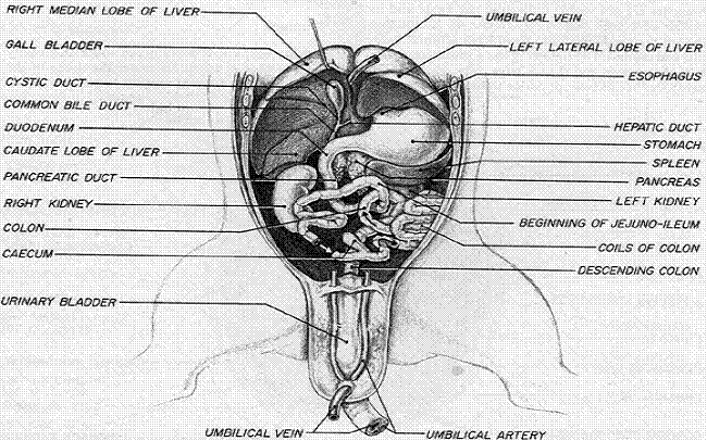
Image: Cow, Eyes and Learning on Pinterest
File Type = .PDF
Credit To @ www.pinterest.com
PDF Download
Open new tab

Image: Sinus Cavity Diagram Sinusitis, Sinus cavities, Sinus
File Type = .PDF
Credit To @ www.pinterest.com
PDF Download
Open new tab
Can I use the push pull pots for the neck or...

Diagram of a cavity. Explore the interactive 3-D diagram. Cavity with a hole. The oral cavity is located just beneath the nasal cavity, the two being separated by the palate [4].Extending from the mouth opening, it continues till above the throat, to the beginning of the oropharynx, the part of the pharynx located just after the oral cavity [5].. Thoracic cavity, also called chest cavity, the second largest hollow space of the body.It is enclosed by the ribs, the vertebral column, and the sternum, or breastbone, and is separated from the abdominal cavity (the body’s largest hollow space) by a muscular and membranous partition, the diaphragm.It contains the lungs, the middle and lower airways—the tracheobronchial tree—the heart.
Vertically it is enclosed by the vertebral column and the abdominal Human body cavity diagram. A chart, plan, or scheme Not to be confused with: Diaphragm – the partition separating the thoracic cavity from the abdominal cavity in mammals;
Houses the spinal cord. It makes up the upper respiratory system along with the paranasal sinuses, oral cavity, pharynx, and larynx [2], and is the. Let us learn in detail about the main parts of the buccal cavity or an oral cavity. A widely used model of a black surface is a small hole in a cavity with walls that are opaque to radiation.
Anterior view of the inferior and superior vena cava in relationship to the ribcage and pelvis. Warms and humidifies the inspired air.; The nasal cavity has four functions: Its upper boundary is the diaphragm, a sheet of muscle and connective tissue that separates it from the chest cavity;
Sep 6, 2014 - oral anatomy diagram | Anatomy of the oral cavity The oral vestibule – It is the slit-like space between the teeth and the buccal cavity and between the lips and cheeks. Sensory cranial nerves help a person to see, smell, and hear. The pulp cavity, sometimes called the pulp chamber, is the space inside the crown that contains the pulp..
Skin Anatomy Diagram Labeled | Diagram Labels {Label Gallery} Get some ideas to make labels for bottles, jars, packages, products, boxes or classroom activities for free. There are four pairs of sinuses (named for the skull bones in which they're located). Thoracic region of the body with the heart and descending aorta prominently visible upon the rib cage and pelvic bones. I'm wondering if any of you have a wiring diagram showing how to do such a thing?
Drains and clears the paranasal sinuses and lacrimal ducts. Cranial cavity–the space occupied by the brain, enclosed by the skull bones. Responsible for sense of smell. And v., thymus in it.
Houses the abdominal cavity and pelvic cavity. As shown in the given diagram, the un-regulated cavity resonator is designed in such a way that two pieces of regulated cavity resonator is joint together with the help of circular waveguide. Diagram Of Heart In Chest Beautiful Body Cavities Diagram Human This is an article covering the boundaries pleural reflections and recesses and pathology related to the pleural cavity. In most cases, the body is described as having two main cavities called the “dorsal and ventral body cavities”.
Diagram synonyms, diagram pronunciation, diagram translation, English dictionary definition of diagram. The spinal cavity is continuous with the cranial cavity. I'm looking to wire up a Lester with a 4 conductor push/pull in the bridge and 2 conductor in the neck. Besides the anterior and posterior apertures, each nasal cavity has a roof, floor, and lateral and medial walls.There are 12 cranial bones in total that contribute to the nasal cavity structure, which include the paired nasal, maxilla, palatine and lacrimal bones, as well as the unpaired ethmoid, sphenoid, frontal and vomer bones.Among all of them, the ethmoid bone is the most important.
It consists of nasal skeleton, which houses the nasal cavity. Some anatomical references do not recognize the dorsal body cavity but we will use it in this example because it’s used by many professionals and colleges. I have a martin 6 strings Jimmy page harness to use. Chest cavity, vena cava:
Abdominal cavity, largest hollow space of the body. Parts of the Buccal. Related For Labeled Diagram Of The Oral Cavity. Spinal cavity–the space occupied by the spinal cord enclosed by the vertebrae column making up the backbone.
Motor cranial nerves help control muscle movements in the head and neck. Removes and traps pathogens and particulate matter from the inspired air. Chest Cavity Chest cavity, aorta: Cavity of the body that is located above the diaphragm.
Diagram Of Oral Cavity Posted on April 16, 2019 by admin Tissue harvesting site and culture medium affect attachment growth phenotype of ex vivo expanded oral mucosal epithelial cells scientific reports human tooth anatomy and physiology diagram oral cavity anatomical diagram of the oropharynx oral cavity labeled diagram of the oral cavity. Its lower boundary is the upper plane of the pelvic cavity. Oral Cavity Anatomy and Structure. In one piece of the regulated cavity resonator input port is made and in the second piece output port is designed.
Body cavities and membranes.

Image: Structure of the Peritoneum and Peritoneal Cavity anatomy
File Type = .PDF
Credit To @ www.pinterest.com
PDF Download
Open new tab
