Diagram Of Kidney Tube

Image: The renal system diagram. Includes both full color and
File Type = .PDF
Credit To @ www.pinterest.com
PDF Download
Open new tab

Image: TTube Placement and Bile Drainage for Liver Transplant
File Type = .PDF
Credit To @ www.pinterest.com
PDF Download
Open new tab

Image: Urinary or excretory system Kidneys, ureters (tubes form
File Type = .PDF
Credit To @ www.pinterest.com
PDF Download
Open new tab
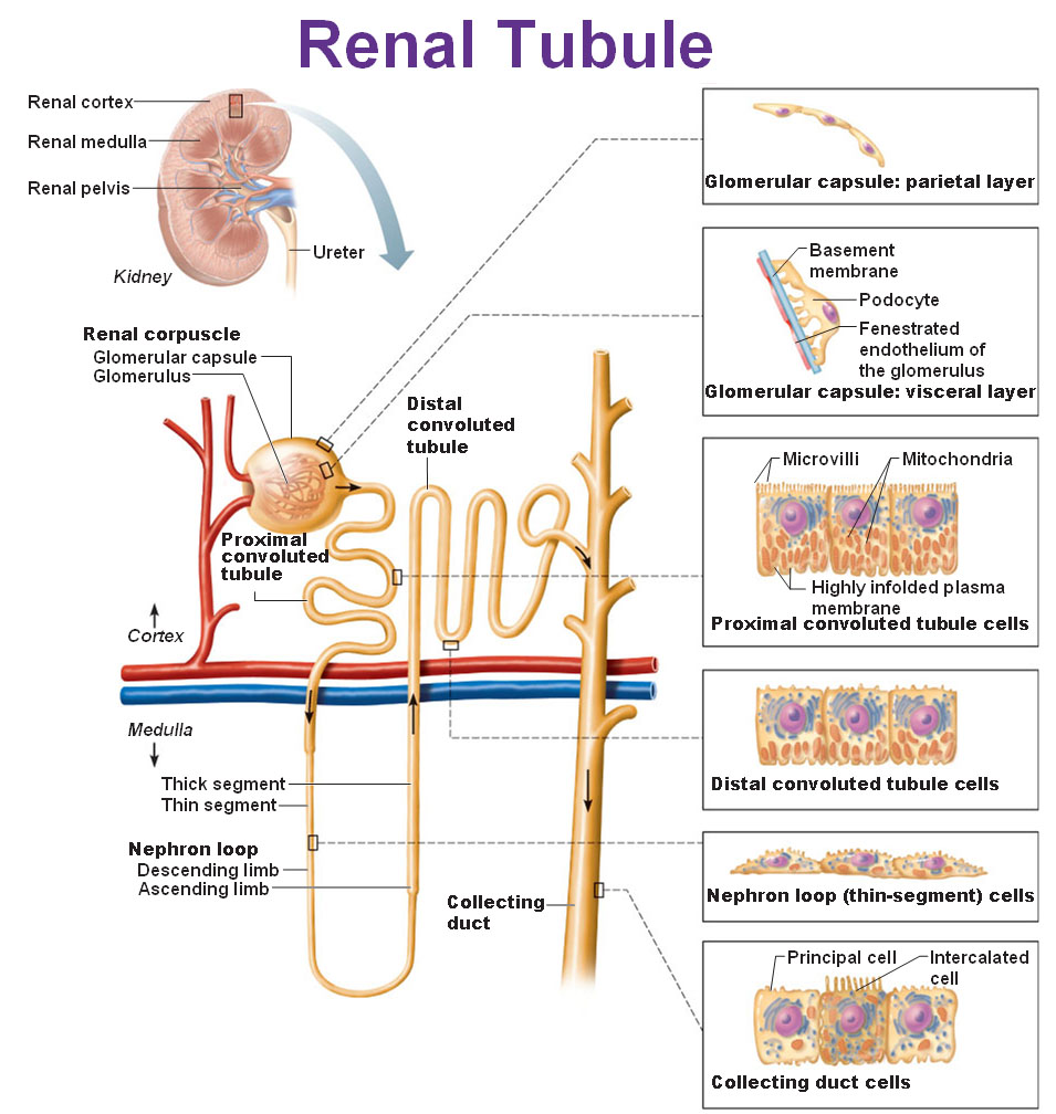
Image: vasa recta Physiology Pinterest Vases, Note and Search
File Type = .PDF
Credit To @ www.pinterest.com
PDF Download
Open new tab

Image: nephron reabsorption secretion diagram Google Search
File Type = .PDF
Credit To @ www.pinterest.com
PDF Download
Open new tab

Image: Label This Diagram Of A Nephron. Study Time
File Type = .PDF
Credit To @ www.pinterest.com
PDF Download
Open new tab
Often called a percutaneous nephrostomy tube, the device attaches to a collection bag that collects and measures urine output.

Diagram of kidney tube. Urinary System Diagram What is a Urinary System Diagram? Diagram Of Kidney Stone Pain 5 out of 5 based on 7 ratings. The second ureter can be normal or only partially developed. Each kidney has around a million tiny.
The functional unit of the kidney is the nephron, of which about one million are found in each kidney. The structure of the urethra (tube) itself is a continuous mucous membrane supported by submucous tissue connecting it to the other structures through which it passes. Kidney pain or flank pain can be acute, relatively constant, and sharp. A thin tube is passed through the urinary tract to the location of the stone.
Ureteral obstruction may be caused by: The tube helps to drain urine from your body. Labeled Diagram of the Human Kidney. A nephrostomy tube is a small rubber tube that is placed through a hole in the skin and that extends into the kidney.The tube allows direct drainage from the kidney.
Each kidney is attached to a ureter, a tube that carries excreted urine to the bladder. The waste and water are excreted as urine. Proximal convoluted tubule (pars convoluta) The pars convoluta (Latin "convoluted part") is the initial convoluted portion.. The kidney is a filter for the blood and works to remove waste materials.
180 L/day is filtered, meaning that it passes through the capillary basement membrane into the glomerular capsules, and passes through the tubules.The rest of the renal blood flow stays inside the capillaries, or instead travels through vascular beds that nourish and support the kidney tissue. The nephron is the structural and functional unit of the kidney. There are about a million nephrons in each kidney. The kidneys also reabsorb and return to the blood needed substances, including amino acids, sugar, sodium, potassium, and other nutrients.The kidneys filter about 200 quarts of blood per day and produce about 2.
The nephron is the basic unit of the kidney. In relation to the morphology of the kidney as a whole, the convoluted segments of the proximal tubules are confined entirely to the renal cortex.. No incisions are made in the body. This is termed "renal colic." This kind of pain is usually seen when a kidney stone or other problem blocks the tube (ureter) that drains the kidney.
However, kidney stones can grow larger in size, even filling the inner hollow structures of the kidney. The urinary system is located directly below the rib cage. Kidney stent (medically called a ureteric stent) is a specially designed hollow tube, made of a flexible plastic material. The two important blood vessels of the kidney are:
Kidney diagram Use this interactive 3-D diagram to explore the kidney. Diagram Of Kidney Stone Pain. That’s the first thought that floated into my brain as Briana Oster, M.D., a former pediatrician who now works at Revitalize Laser Care’s Denver office, talked me through a diagram of the. Nephron (Glomerulus and Tubule) Structure, Diagram, Functions.
The drained urine is collected in a small bag located. The tube allows urine to bypass blocked or damaged ureters in order to avoid the risk of infection or. The renal tubule is the end of the nephron. (The ureter is the tube between the kidney and the bladder.)
Some investigators on the basis of particular functional differences have divided the convoluted part into two segments. The part of the kidney nephron that collects urine and drains into papillary ducts, minor calyx, and major calyx, and finally into the ureter and urinary bladder. Kidney pain is not in itself a diagnosis and could be a symptom of various issues. Each human kidney possesses about 1 -2 millions of nephrons.
Posted on by . The urinary system, at a high level, contains two kidneys, two ureters, a urethra, and a bladder. Each nephron is made up of two main parts: For more anatomy content please follow us and visit our website:
(1) Renal Artery (2. Each human adult kidney contains around 1 million nephrons, while a mouse kidney contains only about 12,500 nephrons. The ureter is the tube through which urine moves from the kidney to the bladder. Urinary system diagrams are illustrations of the urinary system, also referred to as the renal system.
1 litre of blood passes through the kidneys every minute, but not all of that is filtered. It can also control the levels of electrolytes and water that is lost in the urine or retained in the body thereby. A surgeon breaks up the stone and removes the fragments through the tube. A nephrostomy tube is a catheter that’s inserted through your skin and into your kidney.
In addition, they also play an important role in maintaining the water balance of our body. The functional units where the kidney's main functions are performed. In each kidney, there are one million of these structures, called nephrons. (1) Malpighian Body, (2) Renal tubule.
For example, they show that on top of each kidney is an adrenal gland. Kidney stones may start small and not cause any issues at first. However, other processes can cause chronic dull aching with occasionally sharp kidney pain. Waste gets turned into urine, which collects in the kidney's pelvis -- a funnel-shaped structure that drains down a tube called the ureter to the bladder.
Aside from filtration the kidneys also helps with hormone and electrolyte control. It's a long, thin tube that is closed at one end, has two twisted regions interspaced with a long hairpin loop, ends in a long straight portion and is surrounded by capillaries. Some stones stay in the kidney, and will never cause any problems. The mucous coat is continuous with the mucous membrane of the bladder, ureters and kidney.
The ureter is a tube of muscle that pushes urine into the bladder, where it collects and exits the body. They function chiefly to filter blood in order to remove wastes and excess water. Kidney stones can travel down the ureter sometimes. Duplication of the ureter, the tube that carries urine from the kidney to the bladder.
What do the Kidneys do in our body? Each normal human kidney has about 800,000 to one million nephrons, and each nephron has a renal tubule. Renal tubules are part of the nephron, the basic functional unit of the kidney. The main function of the kidney is to filter fluid from the blood and concentrate the solution of waste materials which is passed out as urine.
Nephron is the basic unit of kidney.

Image: Label This Diagram Of A Nephron. Study Time
File Type = .PDF
Credit To @ www.pinterest.com
PDF Download
Open new tab
